
White Paper
Silent Hypoxia Is Not Silent: Blood Rheology, Post-Prandial Inflammation, and Microcirculatory Oxygen Delivery — Insights from ESR, TcPO₂, and Observational Evaluation of XenaGen™
Authors:
Doc X, MD, ScD, MPH, MS
Doc X, RN, BSN, DNP
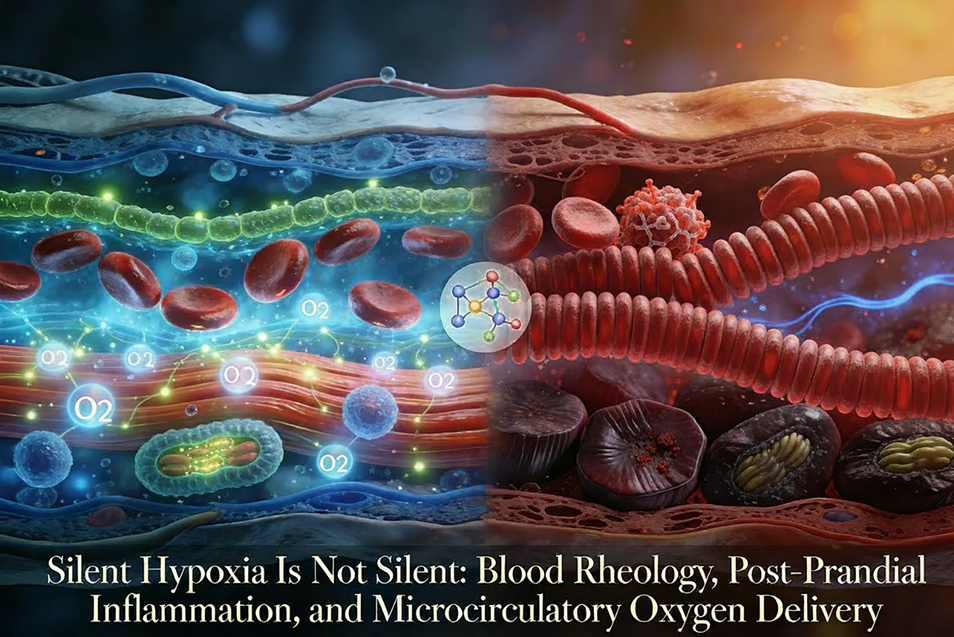
Image 1 Caption:
Post-prandial silent hypoxia vs. enzymatic microcirculatory restoration. This medical illustration demonstrates split-view: Right—post-meal impaired microcirculation with heavy RBC rouleaux stacks (coin-like clumps) and agglutination blocking capillaries, causing implied low skin TcPO₂, faint O₂ trails, hypoxic dark mitochondria, and damaged glycocalyx exposing endothelium; Left—XenaGen™-optimized smooth RBC flow, protected green glycocalyx against inflammation, abundant O₂ diffusion and release to vibrant healthy tissue. Center enzymatic molecules dissolving inflammation-associated fibrin (yellow fibrils).
Abstract
Background:
Oxygenation is routinely assessed using arterial hemoglobin saturation (SpO₂), yet effective tissue oxygen availability is determined downstream at the microcirculatory level by capillary flow dynamics, diffusion distance, glycocalyx integrity, and oxygen release. Clinical observation and tissue-level measurement indicate that oxygen delivery impairment can occur despite preserved SpO₂, particularly during post-prandial inflammatory states. This discordance has contributed to the mischaracterization of clinically meaningful, tissue-level hypoxia as “silent.”
Methods:
This white paper integrates longitudinal physiologic observation, established rheologic principles, inflammatory context reflected by erythrocyte sedimentation rate (ESR), and transcutaneous oxygen tension (TcPO₂) measurement to examine flow- and release-limited tissue hypoxia. Observational evaluation of a mechanism-aligned, enzymatic oxygen-centric wellness adjunct (XenaGen™) is presented within a quality-improvement framework. The analysis emphasizes physiologic plausibility, temporal alignment, and coherence across microcirculatory oxygen delivery, inflammatory burden, and functional manifestations rather than causal inference or comparative efficacy.
Results:
Across observational contexts, post-prandial inflammatory signaling was associated with rheologic shifts consistent with increased plasma viscosity and red blood cell aggregation, sufficient to impair capillary flow despite normal arterial oxygen saturation. TcPO₂ consistently demonstrated post-prandial reductions in tissue oxygen tension that were not detected by saturation-based metrics, revealing a reproducible discordance between systemic oxygen availability and effective tissue-level delivery and release. These tissue-level oxygen constraints aligned temporally with commonly reported functional manifestations, including post-prandial fatigue, reduced alertness, impaired endurance, delayed recovery, and wound-healing inefficiency. Observational TcPO₂ patterns associated with oxygen-centric adjunct use demonstrated internally coherent improvement without exceeding physiologic plausibility or invoking treatment claims.
Conclusions:
Within an observational, hypothesis-generating quality-improvement framework, these findings support reframing so-called “silent hypoxia” as unrecognized, flow- and release-limited tissue hypoxia arising from microcirculatory transport constraints rather than failures of oxygen supply. Normal SpO₂ does not guarantee adequate tissue oxygenation under inflammatory and rheologic stress. TcPO₂, interpreted alongside inflammatory and rheologic context, provides a clinically meaningful window into tissue-level oxygen availability and renders visible physiologic limitations that saturation-based metrics systematically overlook. Oxygen-centric strategies aligned with microcirculatory flow and oxygen release warrant continued structured investigation to better align physiologic assessment with patient experience, recovery dynamics, and tissue-level performance.
Executive Summary
Oxygen delivery impairment is commonly framed as a pulmonary, cardiac, or hematologic failure. Microvascular tissue oxygen availability, however, is ultimately determined downstream—within the microcirculation—by capillary flow, diffusion distance, microvascular glycocalyx integrity, and oxygen release at the tissue interface.
Convergent physiologic observation and tissue-level measurement support an additional, underrecognized mechanism: flow-limited tissue hypoxia driven by dynamic shifts in microvascular transport conditions even when arterial oxygen saturation (SpO₂) remains normal.
Post-prandial inflammatory signaling can acutely increase plasma proteins (including fibrinogen) and alter red blood cell aggregation, producing measurable, transient increases in blood viscosity. Within-individual viscosity rises of approximately 20% relative to baseline have been observed in longitudinal ESR-correlated clinical evaluation and are consistent with rheology literature; changes of this magnitude are sufficient to impair capillary flow and reduce effective tissue oxygen tension and release.
Transcutaneous oxygen tension (TcPO₂) provides a functional physiologic measure of tissue oxygen availability and consistently demonstrates post-prandial reductions in tissue oxygen tension despite preserved SpO₂—highlighting a recurrent discordance between systemic saturation and microvascular tissue-level O₂ delivery and release.
Clinical manifestations frequently labeled as non-specific—including reduced alertness, post-prandial fatigue, delayed recovery, exertional limitation, ischemic symptoms, and impaired wound healing— are physiologically coherent with tissue-level oxygen delivery and release constraints that are not captured by saturation-based metrics.
This paper reframes so-called “silent hypoxia” as unrecognized hypoxia arising from a classification gap between systemic oxygen saturation and tissue-microvascular level oxygen delivery and release. It reviews non-Newtonian rheologic principles, highlights TcPO₂ as a clinically informative endpoint, and evaluates XenaGen™ as a mechanism-aligned wellness bioactive adjunct within an observational, quality-improvement framework focused on microcirculatory flow and oxygen transport dynamics.
Introduction: When “Normal” Masks Physiologic Impairment
Pulse oximetry measures arterial hemoglobin saturation. It does not measure oxygen delivery to tissue.
Clinicians have long understood this distinction in wound care, vascular medicine, and perioperative care, where TcPO₂ is routinely used to assess perfusion and healing potential. What has been underappreciated is how everyday physiologic states—particularly post-prandial inflammation—can impair microvascular tissue oxygenation without triggering conventional alarms.
Individuals report:
- Reduced alertness or cognitive fog
- Post-meal fatigue
- Decreased exercise tolerance
- Delayed post-surgical recovery
- Increased exertional or ischemic symptoms (angina)
When viewed through a saturation-based framework, these manifestations are often mislabeled as non-overt, despite being clinically apparent and physiologically coherent; they remain conceptually under-recognized due to limitations in how tissue oxygen delivery is assessed.
Blood Is Not a Newtonian Fluid
Blood behaves as a non-Newtonian fluid, meaning its viscosity changes dynamically based on:
- Plasma protein concentration (especially fibrinogen)
- Red blood cell aggregation
- Erythrocyte deformability
- Local shear rates within micro vessels
Small rheologic changes that appear modest in bulk measurements produce disproportionately large effects at the capillary level, precisely where oxygen exchange occurs. Oxygen delivery impairment, therefore, can occur without hypoxemia.
ESR, Inflammation, and the Post-Prandial State
Acute-phase reactants characteristically increase during inflammatory signaling, with proportional rises in fibrinogen and related plasma proteins that directly influence blood rheology. The erythrocyte sedimentation rate (ESR) has long served as a non-specific, but clinically practical, integrative marker of this inflammatory burden.
Across more than three decades of ESR-informed clinical observation and longitudinal assessment, a consistent physiologic pattern has emerged:
- Low-grade inflammatory signaling is common rather than exceptional
- Nutrient absorption itself constitutes a normal physiologic inflammatory stimulus
- Meals—particularly those high in fat or rapidly absorbed carbohydrates—are associated with post-prandial increases in IL-6, fibrinogen, and coagulation-related proteins within hours
In isolation, these post-prandial inflammatory responses would be expected to resolve. In practice, however, modern eating patterns characterized by closely spaced meals and snacks frequently result in cumulative inflammatory signaling, producing sustained elevation in plasma viscosity across much of the waking day.
Within this context, longitudinal observation and rheology-supported estimates indicate that, in a subset of individuals, post-prandial blood viscosity may rise by approximately 20% relative to personal baseline. At the microcirculatory level, changes of this magnitude are sufficient to impair capillary flow dynamics and reduce effective tissue oxygen delivery, despite preserved arterial oxygen saturation.
This pattern does not represent disease. Rather, it reflects normal physiologic inflammatory processes expressed at a scale and frequency that meaningfully alter microvascular oxygen transport.
TcPO₂: Making the Invisible Visible
When rheologic changes impair microvascular flow, the resulting oxygen deficit is not adequately captured by saturation-based metrics. Tissue oxygen tension—not arterial saturation—provides the more relevant physiologic signal under these conditions.
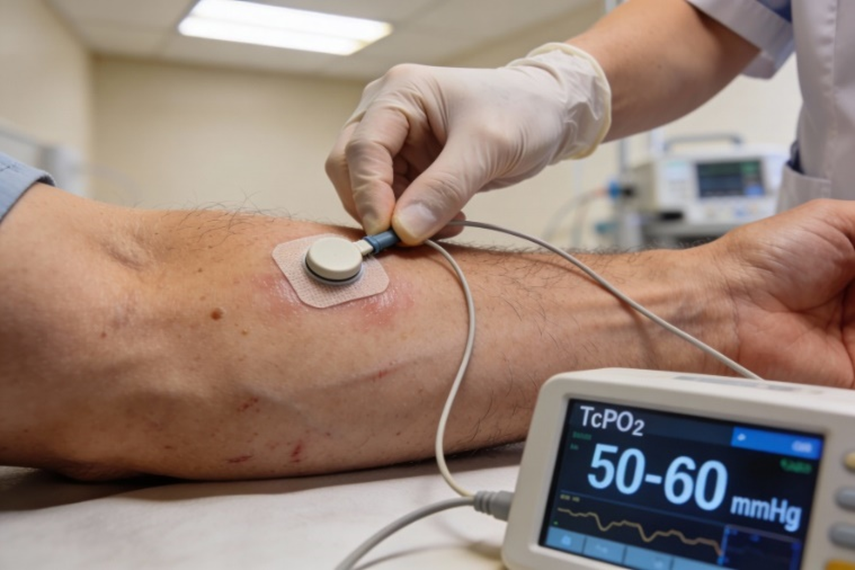
Image 2 Caption
Transcutaneous oxygen measurement (TcPO₂) provides a functional measure of the diffusion of oxygen from capillary beds into surrounding tissue. Unlike pulse oximetry, which measures hemoglobin saturation in large arterial vessels, TcPO₂ integrates the combined effects of perfusion, diffusion distance, capillary flow dynamics, and local metabolic demand. For this reason, TcPO₂ has long been used in wound care, vascular assessment, and perioperative decision-making as a functional indicator of tissue oxygen availability and release.
Within observational and quality-improvement contexts, TcPO₂ consistently demonstrates physiologic phenomena that saturation-based measures fail to detect. Specifically, TcPO₂ reveals post-prandial declines in tissue oxygen tension that occur despite preserved arterial oxygen saturation, highlighting a recurrent discordance between systemic oxygen availability and effective microvascular tissue-level delivery.
Across longitudinal observation, TcPO₂ patterns show:
- Reproducible post-prandial reductions in tissue oxygen tension
- Temporal alignment with inflammatory and rheologic shifts reflected in ESR trends
- Improvement when microcirculatory flow dynamics are favorably altered
These TcPO₂ changes align with functional outcomes—including endurance capacity, wound healing trajectories, and recovery kinetics—supporting its role as a clinically meaningful physiologic endpoint rather than a purely technical or surrogate measurement.
TcPO₂ does not identify a failure of oxygen supply. Rather, it reveals limitations in oxygen transport and release at the microcirculatory level, where capillary flow resistance, red blood cell aggregation, and diffusion constraints converge. In this way, TcPO₂ renders visible a form of flow- and oxygen-release–limited hypoxia that remains concealed when assessment is restricted to arterial saturation alone.
Top of Form
Bottom of Form
Why “Silent Hypoxia” Is a Misnomer
The hypoxia observed in these contexts is not silent. Rather, it is unrecognized.
The term “silent hypoxia” implies an absence of physiologic consequence or patient experience. The reality is the limitation lies not in symptom expression, but in the frameworks traditionally used to assess oxygenation. When evaluation is confined to arterial oxygen saturation, impairments in microvascular tissue-level oxygen delivery and release remain systematically overlooked.
Clinical training and conventional monitoring emphasize indicators such as dyspnea, cyanosis, reduced SpO₂, and gross laboratory abnormalities. When these markers are absent, oxygenation is presumed adequate. This assumption fails to account for flow- and oxygen-release–limited hypoxia arising at the microcirculatory level, where oxygen exchange actually occurs.
Within this gap, patients frequently experience manifestations that are clinically real but conceptually discounted, including:
- Post-prandial fatigue or reduced alertness
- Cognitive slowing or diminished concentration
- Impaired exercise benefits, tolerance, or recovery
- Delayed wound healing
- Wide prediabetic and diabetic random plasma glucose (RPG) level variability
- Exacerbation of exertional or ischemic symptoms
These manifestations do not reflect vague or incidental complaints. They are consistent with reduced tissue oxygen availability and release that is not captured by saturation-based metrics and therefore remains unrecognized within conventional assessment models.
In this context, the descriptor “silent” misattributes the problem. The hypoxia is neither rare nor asymptomatic. It is simply invisible to tools and frameworks optimized for detecting failures of oxygen supply rather than limitations in oxygen transport and release.
Recognizing this distinction reframes oxygen assessment from a binary evaluation of saturation to a physiologic appraisal of delivery, diffusion, release, and utilization. TcPO₂, informed by inflammatory and rheologic context, provides a means to detect this otherwise concealed form of hypoxia and to align observed patient experience with underlying microvascular physiology.
XenaGen™: A Mechanism-Aligned Wellness Intervention
Rationale
XenaGen™ is positioned as a wellness-oriented bioactive adjunct whose enzymatic composition aligns with physiologic processes implicated in post-prandial inflammatory burden, blood rheology, and microcirculatory oxygen delivery and release. Its inclusion within this paper is grounded in mechanistic alignment and physiologic plausibility rather than claims of treatment, prevention, or disease modification.
Within established inflammatory physiology, processes that influence circulating protein burden and post-prandial inflammatory signaling are associated with fibrinogen dynamics, plasma viscosity, red blood cell aggregation, and capillary flow resistance—key determinants of effective tissue oxygen transport and release. These processes provide relevant physiologic context for interpreting tissue-level oxygen measurements during inflammatory states.
Inflammatory signaling is also known to alter endothelial surface properties, including the glycocalyx layer, which can increase diffusion distance and resistance to oxygen release at the capillary–tissue interface. Such effects further contribute to flow- and release-limited tissue hypoxia during inflammatory states.
Within the scope of this paper, XenaGen™ is evaluated solely as a mechanism-aligned adjunct within a quality-improvement, observational context focused on microcirculatory flow and oxygen delivery dynamics.
Related quality-improvement work examining post-surgical wound healing, incorporating transcutaneous oxygen tension (TcPO₂) measurement, has reported internally coherent patterns linking improved tissue oxygen availability and release with more favorable recovery trajectories in observational settings. These findings, detailed separately, provide applied physiologic context that reinforces the oxygen-centric framework described here.18 Additional observational quality-improvement evaluations in populations characterized by metabolic (prediabetes) and vascular (PVD) risk have similarly described favorable directionally aligned, internally coherent patterns when enzymatic oxygen-centric adjuncts were included in patterns of care.19,20
Interpretive Boundaries
Governance & interpretive boundary: This paper is observational and hypothesis-generating within a quality-improvement frame. It reports physiologic measurements and coherent longitudinal patterns without establishing causality. XenaGen™ is discussed as a mechanism-aligned wellness adjunct; observations are presented to clarify microcirculatory oxygen delivery dynamics and to support continued rigorous investigation.
Clinical and Wellness Implications
Unrecognized, flow- and release-limited hypoxia arising from post-prandial inflammatory and rheologic dynamics carries implications across multiple domains of health and performance. Because these effects occur in the presence of preserved arterial oxygen saturation, they remain largely invisible within conventional assessment frameworks, despite their functional relevance.
Within observational contexts, impaired microcirculatory oxygen delivery has been associated with patterns affecting:
- Daytime fatigue and cognitive performance
- Exercise tolerance, endurance, and recovery capacity
- Wound healing trajectories
- Cardiovascular strain during exertion
- Post-operative recovery efficiency
- Cumulative inflammatory burden over time
These implications do not suggest discrete pathology. Rather, they reflect a population-scale physiologic pattern in which every day inflammatory responses, when expressed with sufficient frequency and magnitude, alter oxygen transport at the tissue level. The clinical significance lies not in the rarity of the phenomenon, but in its persistence and invisibility when assessment is restricted to saturation-based metrics.
By reframing oxygen impairment as a matter of delivery and release rather than supply alone, these observations expand the interpretive context for commonly reported functional complaints that are otherwise dismissed or compartmentalized.
Practical Considerations for Clinicians
For clinicians, these observations reinforce several practical principles relevant to everyday care and quality-improvement contexts.
First, normal arterial oxygen saturation does not guarantee adequate tissue oxygenation. Inflammatory burden, rheologic shifts, and microcirculatory flow resistance materially influence oxygen delivery downstream of the arterial compartment.
Second, inflammatory markers such as ESR can retain interpretive value even when results fall within conventional reference ranges. Longitudinal trends and contextual timing—particularly in relation to meals, recovery, and exertion—may be more informative than isolated measurements.
Third, transcutaneous oxygen tension (TcPO₂) offers a clinically meaningful window into tissue-level oxygen availability, particularly in settings where functional limitation, impaired healing, or recovery inefficiency is observed despite otherwise reassuring systemic metrics.
Finally, physiologic variables including meal composition, meal timing, physical activity, and recovery states influence rheologic load and oxygen transport. Mechanism-aligned, non-pharmacologic strategies—including enzyme-based wellness adjuncts such as XenaGen™—may support physiologic resilience within observational and quality-improvement frameworks when applied with appropriate boundaries and expectations.
These considerations do not constitute diagnostic guidance or therapeutic recommendation. Rather, they highlight opportunities to align physiologic assessment more closely with patient experience and microvascular function.
Conclusion
Across longitudinal observation, ESR-informed inflammatory context and TcPO₂ measurement converge on a coherent physiologic pattern. Within this framework, post-prandial rheologic shifts associated with inflammation reduce tissue oxygen tension despite preserved SpO₂. This framework aligns symptom patterns—post-prandial fatigue, reduced alertness, impaired endurance, delayed recovery, and wound-healing inefficiency—with measurable tissue-level oxygen constraints rather than with incidental or benign complaints.
Within an observational, quality-improvement frame, XenaGen™ is mechanism-aligned with upstream processes influencing post-prandial rheology and microvascular flow. Observed TcPO₂ improvements during post-prandial and recovery states support physiologic coherence without extending to causal or efficacy claims.
What has been described as “silent hypoxia” is not an absence of physiology. It is a detection gap: arterial saturation can remain normal while microcirculatory transport conditions constrain tissue oxygen delivery and release.
Seeing oxygen clearly requires assessment beyond hemoglobin saturation alone. TcPO₂, interpreted alongside inflammatory and rheologic context, provides a stable physiologic foundation for continued investigation into oxygen-centric strategies that support resilience, recovery kinetics, and tissue-level performance.
References
Postprandial Inflammation & Rheology/ESR (5 refs)
Sun Y, Han L, et al. Blood viscosity in type 2 diabetes: role of fibrinogen & RBC aggregation. Front Physiol. 2022;13:827428. doi:10.3389/fphys.2022.827428
Heresi GA, et al. IL-6 & fibrinogen variants in postprandial inflammation & CVD risk. PLoS One. 2011;6(12):e50212. doi:10.1371/journal.pone.00289374
Brunner H. Blood viscosity & hypertension: fibrinogen’s role. J Hypertens. 2007;25(4):647-651. doi:10.1097/HJH.0b013e328014ca78
Larsson PT, et al. Postprandial IL-6 & fibrinogen rise after swine dust inhalation. Am J Respir Crit Care Med. 1999;160(3):919-925. doi:10.1164/ajrccm.160.3.9809055
Lowe GDO, et al. Fibrinogen, viscosity & CVD risk post-meal. Mol Nutr Food Res. 2020;64(7):1901035. doi:10.1002/mnfr.201901035
Silent/Tissue Hypoxia & Microcirculation (5 refs)
Umbrello M, Formenti P. Silent hypoxia in COVID-19: RBC NO & vasodilation despite low O2. Open Biol. 2020;10(10):200181. doi:10.1098/rsob.200181
Mayrovitz HN. Microcirculation in wound healing: O2 delivery & glycocalyx. In Wound Healing (2023): Ch. 10. Humana Press. doi:10.1007/978-3-031-33081-5_10
Botek M, Krejčířová K. Glycocalyx degradation & endothelial dysfunction in hypoxia. Front Physiol. 2022;13:827428. doi:10.3389/fphys.2022.827428
Sen CK. Oxygen implications for wound healing: diffusion & release limits. Wound Repair Regen. 2012;20(1):1-18. doi:10.1111/j.1524-475X.2011.00743.x
Sheffield PJ. TcPO2 in hyperbaric O2 & tissue perfusion. Undersea Hyperb Med. 2015;42(5):413-428. PMID:26453415
TcPO2, ESR & Clinical Discordance (5 refs)
Bordonado-Murcia A, et al. TcPO2 prognostic in diabetic ulcers: systematic review protocol. Diagnostics (Basel). 2025;15(7):909. doi:10.3390/diagnostics15070909
Monsen RE. TcPO2 clinimetrics & chronic wound healing. Wound Repair Regen. 2014;22(5):583-590. doi:10.1111/wrr.12209
Brigden ML. ESR clinical utility in inflammation/rheology. Am Fam Physician. 1999;60(5):1443-1450. PMID:10524488
Ridker PM, et al. IL-6 receptor inhibition & vascular inflammation (ESR/fibrinogen). Eur Heart J. 2015;36(8):482-490. doi:10.1093/eurheartj/ehu459
Schreml S, et al. Oxygen in acute/chronic wounds: TcPO2 discordance. Br J Dermatol. 2010;163(2):257-268. doi:10.1111/j.1365-2133.2010.09867.x
Xenakis, A. R-S. X., & Xenakis, A. X. (2025). Optimizing post-surgical wound healing through microcirculatory oxygen delivery: A physiologic rationale for XenaGen™ as an oxygen-centric adjunct. XenaDigm. https://xenadigm.com/qi-white-papers-for-xenagen/
Xenakis, A. R-S. X., & Xenakis, A. X. (2025). Prediabetes quality-improvement study using an oxygen-centric adjunct as intervention: XenaGen™ clinical QI observational analysis. XenaDigm. https://xenadigm.com/qi-white-papers-for-xenagen/
Xenakis, A. R-S. X., & Xenakis, A. X. (2025). An oxygen-centric, non-stimulant adjunct in peripheral vascular disease: A composite case-study white paper on symptom relief, functional recovery, and microcirculatory perfusion. XenaDigm. https://xenadigm.com/qi-white-papers-for-xenagen/