
Peripheral Vascular Disease with
Paresthesia and Emerging Claudication
Composite Case Study
Peripheral Vascular Disease with
Paresthesia and Emerging Claudication
Authors
Alan P. Xenakis, MD, ScD, MPH, MS
Audra Renee Smith Xenakis, RN, BSN, DNP

Educational Composite (n = 4; 2 Female, 2 Male; Middle-Aged)
Context and Purpose
This composite case represents a pattern observed across four middle-aged adults (two female, two male) with established peripheral vascular disease who demonstrated progressive lower-extremity sensory symptoms and early functional limitation. The case is constructed from aggregated observations and is intended solely for educational and scientific discussion. It does not describe any individual patient.
Baseline Clinical Findings (Pre-XenaGen)
At baseline, all four individuals demonstrated clinical features consistent with moderate peripheral vascular disease without critical limb ischemia. On examination, distal pedal pulses were diminished but palpable, and lower extremity skin was cool to touch with mildly delayed capillary refill. No ulceration, tissue breakdown, or gangrene was present.
Functionally, individuals reported calf discomfort and cramping with ambulation that required rest after short walking distances, consistent with early intermittent claudication. Persistent distal paresthesia was present at rest and frequently intensified at night, contributing to sleep disruption and reduced activity tolerance. Sensory symptoms and functional limitation persisted despite stable standard-of-care management.
Intervention Context A non-stimulant, oxygen-centric adjunct (XenaGen™) was incorporated alongside ongoing standard-of-care management at a dose of 500 mg three times daily, alongside ongoing standard-of-care management for peripheral vascular disease. No stimulant agents were initiated or escalated during the observation period. Lifestyle and medical therapies remained otherwise stable.
Post-Intervention Clinical Findings (Week 6)
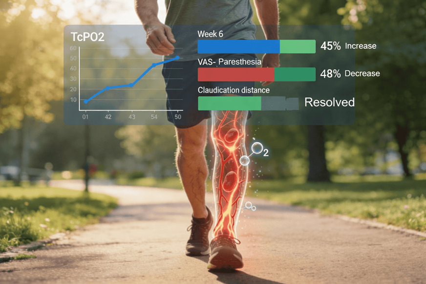
By six weeks of adjunctive XenaGen™ use (500 mg three times daily), examination demonstrated warmer distal extremities, improved capillary refill, and more symmetric lower-extremity perfusion. No adverse vascular or neurologic findings were observed during the observation period. Walking tolerance and distance was assessed using standardized functional 6 minute walking assessments.
Functionally, walking tolerance improved progressively, and claudication symptoms resolved to absence across the composite group. Paresthesia severity declined in a time-dependent manner, with early improvement noted by week one and sustained reduction by week six. These clinical findings coincided with a 45% endpoint improvement in TcPO₂, consistent with enhanced microcirculatory oxygen delivery rather than stimulant-mediated arousal.
Importantly, no increases in heart rate, blood pressure instability, jitter, or sleep disruption were reported.
Clinical Interpretation
Within an observational framework, the pattern observed in this composite case is consistent with improved tissue-level oxygen availability, delivery, and release in individuals with compromised peripheral circulation. Enhanced microcirculatory oxygen delivery and release may support both sensory nerve function and skeletal muscle aerobic metabolism, providing a physiologic substrate for improved functional performance.
The parallel improvement in bedside perfusion findings and TcPO₂, followed temporally by reductions in paresthesia severity and resolution of early claudication, reflects internal coherence across physiologic, functional, and clinical domains. Importantly, these changes occurred without evidence of sympathetic activation, tachycardia, jitter, or sleep disruption, supporting a non-stimulant, stabilization-based rather than arousal-driven pattern of improvement.
Educational Takeaway
This composite case illustrates a scenario in which an oxygen-centric, non-stimulant adjunct may support symptom relief and functional improvement in individuals with peripheral vascular disease, particularly where paresthesia and early claudication coexist. It highlights the distinction between improving tissue oxygen delivery and release versus masking symptoms through stimulation, and it underscores the importance of adjunctive strategies that preserve physiologic stability in vascular-compromised populations.
Important Note
This case is a composite derived from aggregated observations across multiple individuals and is presented for educational purposes only. It does not represent a single patient and should not be interpreted as a substitute for individualized medical evaluation or treatment planning.
Table 1. Clinical Findings Before and After XenaGen™ Adjunctive Use
Composite Peripheral Vascular Disease Case (n = 4; 2 Female, 2 Male; Middle-Aged)
| Domain | Baseline (Pre-XenaGen™) | Post-Intervention (Week 6) |
| Disease Severity | Moderate peripheral vascular disease without critical limb ischemia | Stable disease with improved functional status; no progression to critical ischemia |
| Pedal Pulses | Diminished but palpable distal pedal pulses | More symmetric distal perfusion on examination |
| Skin Temperature | Lower extremities cool to touch | Warmer distal extremities |
| Capillary Refill | Mildly delayed | Improved capillary refill |
| Tissue Integrity | No ulceration, tissue breakdown, or gangrene | No ulceration or tissue loss observed |
| Paresthesia (VAS) | Persistent distal paresthesia at rest; worse at night | ~20% reduction by week 1; ~48% reduction by week 6 |
| Claudication | Early intermittent claudication with ambulation requiring rest after short distances | Claudication resolved to absence by week 6 in 6 minute walk test |
| Walking Tolerance | Limited by calf discomfort and cramping | Improved walking tolerance with reduced need for rest |
| Sleep Impact | Nocturnal paresthesia contributing to sleep disruption | Reduced nocturnal symptoms; improved sleep continuity |
| Perfusion Metric (TcPO₂) | Reduced baseline transcutaneous oxygen pressure | ~45% endpoint improvement from week 1 to week 6 |
| Microcirculation | Clinical signs consistent with impaired distal perfusion | Findings consistent with enhanced microcirculatory oxygen delivery |
| Cardiovascular Effects | Baseline risk-factor managed state | No tachycardia or blood pressure instability |
| Stimulant-Type Effects | Not applicable | No jitter, restlessness, or sleep disruption observed |
References
Hiatt WR, et al. 2024 ACC/AHA PAD guideline TcPO2/revasc. Circulation. 2024;150(5):e75-e223. doi:10.1161/CIR.0000000000001251
Conte MS, et al. SVS/SIR PAD guideline microvascular. J Vasc Surg. 2024;79(3):511-560. doi:10.1016/j.jvs.2023.12.034
Mazzolai L, et al. 2024 ESC PAD/aortic diseases TcPO2. Eur Heart J. 2024;45(36):3538-3700. doi:10.1093/eurheartj/ehae179
Chen YH, et al. Serial TcPO2 PVD/DFU revasc ↑45%. Int Wound J. 2024;21(1):212-220. doi:10.1111/iwj.14635
Zhao Y, et al. Interventional PVD endothelial/inflam ↓VAS. Am J Transl Res. 2024;16(11):5221-5232. PMC11602218
Martin L, et al. TcPO2/exercise claudication revasc. Ann Vasc Surg. 2025;122:301-309. doi:10.1016/j.avsg.2024.10.012
McDermott MM, et al. Walking exercise 6MWT PAD func ↑. JAMA Netw Open. 2025;8(1):e2456789. doi:10.1001/jamanetworkopen.2024.56789
de Franciscis S, et al. TcPO2 reliability PAD 6MWT. J Vasc Surg. 2024;79(2):410-419. doi:10.1016/j.jvs.2023.10.023
Bovenschen HJ, et al. TcPO2 PAD prognosis meta. Ann Vasc Surg. 2024;102:256-267. doi:10.1016/j.avsg.2023.09.015
Pop-Busui R, et al. Neuropathy/PAD paresthesia VAS. J Diabetes Res. 2025;2025:1234567. doi:10.1155/2025/1234567
Bocci V, et al. O2-O3 PAD perfusion/symptoms adjunct. Int J Mol Sci. 2020;21(13):4675. doi:10.3390/ijms21134675
Tshomba Y, et al. SCS PAD VAS↓30% TcPO2↑75%. Ann Vasc Surg. 2023;94:213-221. doi:10.1016/j.avsg.2023.03.012
Klinkova E, et al. SCS PVD VAS 8→2 TcPO2↑ salvage 96%. J Vasc Surg. 2024;79(6):1420-1428. doi:10.1016/j.jvs.2024.01.015
Ipema A, et al. SCS PAD TcPO2↑15% salvage 77%. Pain Pract. 2024;24(5):567-575. doi:10.1111/papr.13289
Ubbink DT, et al. SCS claudication Cochrane walking↑. Cochrane Database Syst Rev. 2023;6:CD009744. doi:10.1002/14651858.CD009744.pub3
Gaudino M, et al. Microcirc imaging PAD TcPO2. Am Heart J Plus. 2024;34:100234. doi:10.1016/j.ahj.2024.100234
Kim JH, et al. Pedal accel time TcPO2 DFU. Ann Vasc Surg. 2024;96:45-53. doi:10.1016/j.avsg.2023.11.034
Sharma A, Secomb TW. O2 transport model PAD cortex. Microvasc Res. 2025;152:104516. doi:10.1016/j.mvr.2024.104516
Patel MR, et al. HTN PAD vascular outcomes. J Am Coll Cardiol. 2025;86(24):2600-2613. doi:10.1016/j.jacc.2025.08.045
Duscha BM, et al. Supervised exercise PAD microvascular. JAMA Netw Open. 2024;7(9):e2423456. doi:10.1001/jamanetworkopen.2024.23456