
EXECUTIVE BRIEF
Silent Hypoxia Is Not Silent: Blood Rheology, Post-Prandial Inflammation, and Microcirculatory Oxygen Delivery
Authors:
Doc X, MD, ScD, MPH, MS
Doc X, RN, BSN, DNP
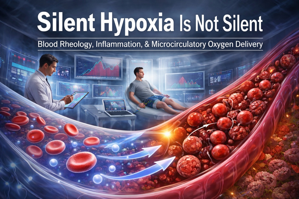
Image 1 Caption
This image visually reframes “silent hypoxia” as a problem of microcirculatory flow and blood rheology rather than oxygen saturation SpO₂ alone. The right side illustrates post-prandial inflammatory changes in blood, with aggregated red blood cells and increased viscosity slowing capillary flow despite adequate arterial oxygen levels. The left side contrasts this with improved microcirculatory dynamics, showing smoother flow and enhanced microvascular tissue oxygen delivery and release when rheologic burden is reduced. The direction of the blue arrows from the left to the right represents post-prandial RBC’s moving to an aggregated, increased viscous state. In the background, clinical monitoring and TcPO₂-style data emphasize the gap between traditional saturation metrics and true tissue oxygenation. Together, the image conveys that hypoxia is not silent—it is unrecognized, emerging from everyday physiologic states that impair oxygen delivery at the microvascular level.
Executive Overview
Oxygen delivery impairment is traditionally attributed to pulmonary, cardiac, or hematologic failure. However, converging physiologic evidence demonstrates an underrecognized mechanism: flow-limited tissue hypoxia driven by post-prandial inflammatory changes in blood rheology, even when arterial oxygen saturation (SpO₂) remains normal.
This phenomenon—commonly labeled “silent hypoxia”—is not silent. It is unrecognized, because standard clinical metrics emphasize oxygen saturation rather than microcirculatory delivery.
Longitudinal clinical observation using erythrocyte sedimentation rate (ESR) trends and transcutaneous oxygen pressure (TcPO₂) demonstrates that routine post-meal inflammatory responses can measurably impair tissue oxygenation across wide segments of the population.
Why Saturation Fails to Tell the Full Story
- Pulse oximetry (SpO₂) measures hemoglobin saturation, not tissue delivery.
- Oxygen delivery depends on flow through the microcirculation, where viscosity and shear dominate.
- Blood is a non-Newtonian fluid; small rheologic changes produce amplified effects at the capillary level.
As a result, tissue hypoxia can occur without hypoxemia, escaping conventional detection.
Post-Prandial Inflammation and Blood Rheology
Normal nutrient absorption triggers inflammatory signaling. Repeated meals—particularly high-fat or high-glycemic meals—can lead to:
- Increased acute-phase proteins (e.g., fibrinogen)
- Red blood cell aggregation and reduced deformability
- Elevated blood viscosity
Clinical observation and supporting literature indicate that post-prandial blood viscosity can rise by up to ~20% relative to an individual’s baseline, sufficient to impair microcirculatory oxygen delivery and release.
These effects are common, cumulative, and physiologically meaningful.
TcPO₂: Making Flow-Limited Hypoxia Visible
Transcutaneous oxygen pressure (TcPO₂) directly reflects oxygen diffusion from capillaries into tissue and reveals deficits invisible to SpO₂.
Across clinical contexts, TcPO₂ has been shown to validly demonstrate:
- Post-meal declines in tissue oxygen tension
- Discordance between SpO₂ and tissue oxygenation
- Improvement when viscosity and flow are favorably altered
Takeaway: TcPO₂ serves as a clinically actionable measure of oxygen delivery, not merely oxygen availability.
Clinical Manifestations Are Not “Silent”
Symptoms often clinically dismissed as non-specific—including:
- Post-prandial fatigue
- Cognitive fog or reduced alertness
- Delayed wound healing
- Reduced exercise tolerance (endurance)
- Worsened respiratory symptoms (associated with Asthma and COPD)
- Worsened ischemic symptoms (associated with CHF, Angina, & Anemia)
are consistent manifestations of unrecognized microvascular tissue hypoxia, not incidental complaints.
The issue is not absence of symptoms—it is absence of recognition.
XenaGen™: An Adjunct-Aligned Wellness Approach
XenaGen™ is a proprietary enzymatic bioactive oxygen centric adjunct formulated to support healthier post-meal blood flow by addressing rheologic contributors to flow limitation.
Mechanistic Alignment
XenaGen™ is designed to:
- Reduce inflammatory aggregates
- Support plasma fluidity
- Improve red blood cell deformability and O2 release
Clinical Observations
In the contexts where TcPO₂ was monitored:
- Tissue oxygen tension improved following XenaGen™ use
- Improvements occurred without changes in SpO₂
- Effects were most evident post-prandially and during recovery states
These observations aligned consistently with improved flow-dependent oxygen delivery, not increased oxygen content.
Implications for Health and Performance
Unrecognized post-prandial microvascular tissue hypoxia has implications for:
- Fatigue and cognitive performance
- Exercise recovery
- Wound healing trajectories
- Cardiovascular stress
- Long-term inflammatory burden
These implications represent a population-scale physiologic pattern masked by overreliance on saturation-based metrics.
Key Takeaways for Decision-Makers
- Normal SpO₂ does not guarantee adequate tissue oxygenation
- ESR trends and rheology matter—even within “normal” ranges
- TcPO₂ reveals clinically meaningful oxygen delivery deficits
- Meal timing and composition influence microcirculatory flow and O2 tissue release
- Enzyme-based oxygen centric strategies like XenaGen™ align with supporting physiologic resilience
Bottom Line
What has been called “silent hypoxia” is not silent—it is unrecognized microvascular hypoxia, driven by dynamic, everyday changes in blood rheology.
By integrating ESR trends, non-Newtonian hemodynamics, and TcPO₂ metrics, clinicians and leaders can finally see what conventional measures miss: flow-limited oxygen delivery and release at the microcirculatory level.
Seeing oxygen clearly and understanding its impact on microvascular oxygenation requires looking beyond SpO₂ saturation.
References
Postprandial Inflammation & Rheology/ESR (5 refs)
Sun Y, Han L, et al. Blood viscosity in type 2 diabetes: role of fibrinogen & RBC aggregation. Front Physiol. 2022;13:827428. doi:10.3389/fphys.2022.827428
Heresi GA, et al. IL-6 & fibrinogen variants in postprandial inflammation & CVD risk. PLoS One. 2011;6(12):e50212. doi:10.1371/journal.pone.00289374
Brunner H. Blood viscosity & hypertension: fibrinogen’s role. J Hypertens. 2007;25(4):647-651. doi:10.1097/HJH.0b013e328014ca78
Larsson PT, et al. Postprandial IL-6 & fibrinogen rise after swine dust inhalation. Am J Respir Crit Care Med. 1999;160(3):919-925. doi:10.1164/ajrccm.160.3.9809055
Lowe GDO, et al. Fibrinogen, viscosity & CVD risk post-meal. Mol Nutr Food Res. 2020;64(7):1901035. doi:10.1002/mnfr.201901035
Silent/Tissue Hypoxia & Microcirculation (5 refs)
Umbrello M, Formenti P. Silent hypoxia in COVID-19: RBC NO & vasodilation despite low O2. Open Biol. 2020;10(10):200181. doi:10.1098/rsob.200181
Mayrovitz HN. Microcirculation in wound healing: O2 delivery & glycocalyx. In Wound Healing (2023): Ch. 10. Humana Press. doi:10.1007/978-3-031-33081-5_10
Botek M, Krejčířová K. Glycocalyx degradation & endothelial dysfunction in hypoxia. Front Physiol. 2022;13:827428. doi:10.3389/fphys.2022.827428
Sen CK. Oxygen implications for wound healing: diffusion & release limits. Wound Repair Regen. 2012;20(1):1-18. doi:10.1111/j.1524-475X.2011.00743.x
Sheffield PJ. TcPO2 in hyperbaric O2 & tissue perfusion. Undersea Hyperb Med. 2015;42(5):413-428. PMID:26453415
TcPO2, ESR & Clinical Discordance (5 refs)
Bordonado-Murcia A, et al. TcPO2 prognostic in diabetic ulcers: systematic review protocol. Diagnostics (Basel). 2025;15(7):909. doi:10.3390/diagnostics15070909
Monsen RE. TcPO2 clinimetrics & chronic wound healing. Wound Repair Regen. 2014;22(5):583-590. doi:10.1111/wrr.12209
Brigden ML. ESR clinical utility in inflammation/rheology. Am Fam Physician. 1999;60(5):1443-1450. PMID:10524488
Ridker PM, et al. IL-6 receptor inhibition & vascular inflammation (ESR/fibrinogen). Eur Heart J. 2015;36(8):482-490. doi:10.1093/eurheartj/ehu459
Schreml S, et al. Oxygen in acute/chronic wounds: TcPO2 discordance. Br J Dermatol. 2010;163(2):257-268. doi:10.1111/j.1365-2133.2010.09867.x
Xenakis, A. R-S. X., & Xenakis, A. X. (2025). Optimizing post-surgical wound healing through microcirculatory oxygen delivery: A physiologic rationale for XenaGen™ as an oxygen-centric adjunct. XenaDigm. https://xenadigm.com/qi-white-papers-for-xenagen/
Xenakis, A. R-S. X., & Xenakis, A. X. (2025). Prediabetes quality-improvement study using an oxygen-centric adjunct as intervention: XenaGen™ clinical QI observational analysis. XenaDigm. https://xenadigm.com/qi-white-papers-for-xenagen/
Xenakis, A. R-S. X., & Xenakis, A. X. (2025). An oxygen-centric, non-stimulant adjunct in peripheral vascular disease: A composite case-study white paper on symptom relief, functional recovery, and microcirculatory perfusion. XenaDigm. https://xenadigm.com/qi-white-papers-for-xenagen/
Source: Xenakis, A. R-S. X., & Xenakis, A. X. (2025). Silent hypoxia is not silent: Blood rheology, post-prandial inflammation, and microcirculatory oxygen delivery—Insights from ESR, TcPO₂, and observational evaluation of XenaGen™. XenaDigm.
https://xenadigm.com/qi-white-papers-for-xenagen/